Introduction
- Learn
- Try It
- Task
- Unit Vocab
- Course Vocab
Introduction
In the last lesson, we learned that there are 3 systems of the heart: plumbing, electrical, and mechanical. We discussed in depth the plumbing and mechanical systems. In this lesson, we will discuss the electrical system of the heart.
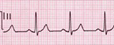
Image 1
Image 1 is an ECG tracing of the heart ("electrical system"). This is the electrical activity occuring in the heart to create contractions of the heart muscle.
Notice the two ECG images below. What is different between the two?
The first image is a normal ECG of a healthy heart. The second image is a heart in trouble and not able to pump blood out to the body. This heart would need to be shocked.
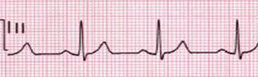
Image 1 Normal Rhythm of the Heart
Image 2 Abnormal Rhythm called V-Tach
Now, let's learn more about the heart and its electrical system.
| Lesson Objectives |
|
Following successful completion of this lesson, students will be able to...
Enduring Understandings
The above objectives correspond with the Alabama Course of Study:Anatomy and Physiology standards: 7a and 7b |
![]()