Learn

Overview of the Heart
Review the Overview of the Heart presentation below. PBS login information.
Overview of the Heart fullscreen version | Overview of the Heart text version
Note: The presentation may take a moment to load.
Mechanical System
Chambers of the Heart
The heart is composed of 4 chambers where blood travels through to be pumped to the lungs and body.
- Right Atrium
- Right Ventricle
- Left Atrium
- Left Ventricle
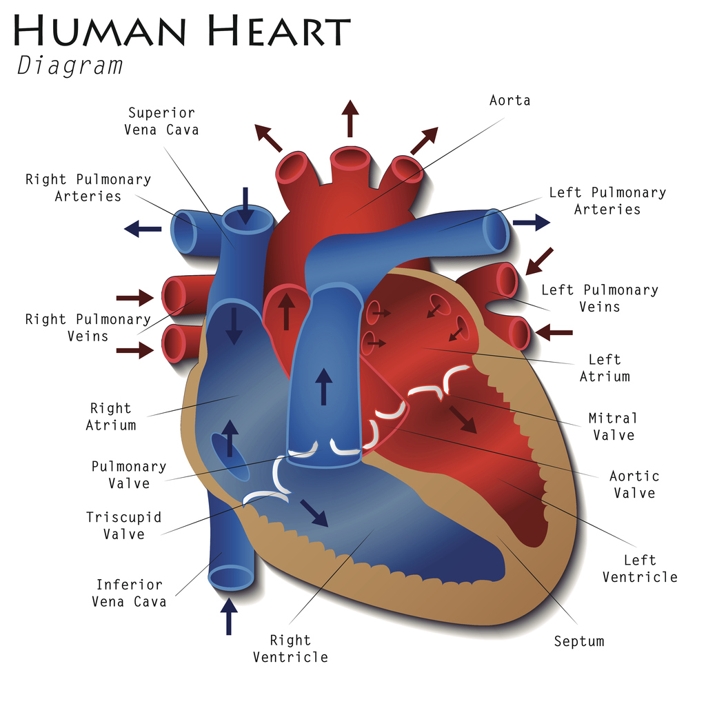
Image 9: Diagram of the Human Heart
Open larger version of Image 9 here.
Valves of the Heart
Here you see an image showing the 4 valves of the heart. They function to maintain a one-way direction of blood flow.
- Atrioventricular (AV) Valves – those valves found between the atria and ventricles
- Tricuspid Valve – on the right side of the heart
- Mitral Valve – on the left side of the heart
- Semilunar Valves – those valves at the top of the heart
- Pulmonary Valve – on the right side of the heart
- Aortic Valve – on the left side of the heart
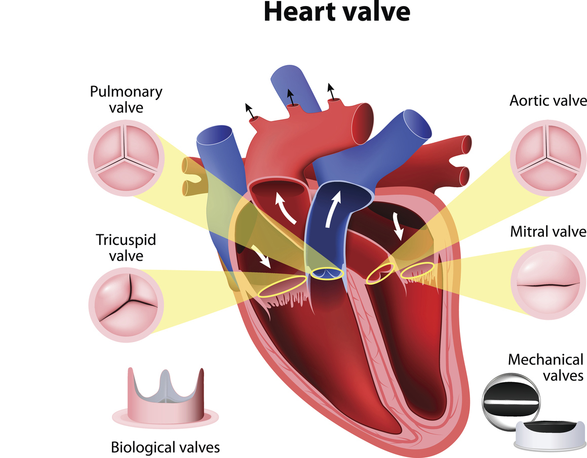
Image 10: Heart Valves
Open larger version of Image 10 here.
![]() Check your understanding...
Check your understanding...
Which 2 chambers are found at the top of the heart (or superior)?
What are the names of the 4 chambers of the heart?
Which valve prevents blood from back-flowing into the left atrium?
Can you identify the number representing the right atrium in the diagram below (Image 11)?

Image 11: Heart Valves
Open larger version of Image 11 here.
Cardiac Cycles
The muscular contractions of the heart occur in repeating sequences of mechanical and electrical events. This repeating sequence is called a cardiac cycle, and this cycle can be divided into 2 phases:
- Systole Phase - period of contraction that the heart undergoes while it pumps blood into circulation
- Diastole Phase - period of relaxation that occurs as the chambers fill with blood
View Mayo Clinic: Heart and circulatory system (03:04) video.
Cardiac cycle details:
- When the heart beatsthe top chambers (atria) contract simultaneously, blood flows through open AV valves to fill the ventricles with blood.
- The AV valves close after blood is emptied from the atria (ventricular diastole phase).
- The bottom chambers (ventricles) are filled with blood and contract simultaneously, and blood flows out to the lungs and body through open semilunar valves (ventricular systole phase).
- The semilunar valves close after blood is emptied from the ventricles.
The cardiac cycle repeats at a resting adult heart rate of 60-80 beats per minute. Open Cardiac Cycle animation to see this process.
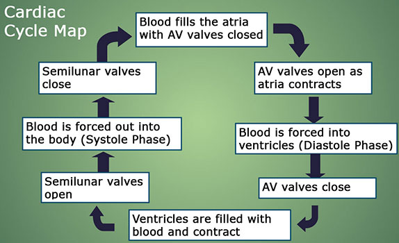
Image 12: The Cardiac Cycle
Open larger version of Image 12 here.
Sounds of a Heartbeat
The heart makes sounds as it beats and these sounds can be heard using an instrument called a stethoscope (seen in the image below).

Image 13: A doctor listening to a baby's heartbeat with a stethoscope.
Listening to the heart for sounds with a stethoscope is called auscultation. The sounds it makes are a "Lub-Dub. These sounds are caused form the turbulent blood flow and closing of the heart valves.
- The "Lub" occurs when the tricuspid and mitral valves close.
- The "Dub" occurs when the pulmonary and aortic valves close.
Now, let's listen to heart sounds that are normal and abnormal.
Abnormal heart sounds - a murmur
- You will hear a "swooshing" sound. The "swooshing" sound is caused by turbulent blood back-flowing through a valve. In this case, it is the mitral valve.
- You can learn more about heart valve problems at the American Heart Association website.
![]() Check your understanding...
Check your understanding...
The cardiac cycle consists of a distinct relaxation and contraction phase. Which term is typically used to refer to ventricular contraction? Systole or Diastole
What is missing in the concept map below of the cardiac cycle (image below)?
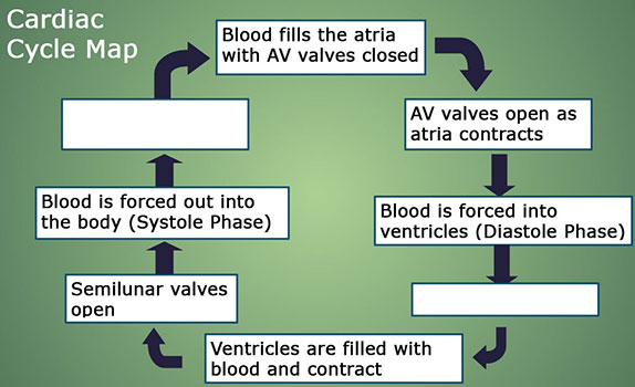
Image 14: The Cardiac Cycle with parts missing
Open larger version of Image 14 here.
What produces the "dub" sound when auscultating the heart?
What can cause the "swooshing" abnormal heart sound of a murmur as heard in this lesson?
Plumbing System
Blood Flow and Volume
The heart pumps about all of the body's blood volume (5 liters) through the body in one minute. Blood volume measurements can be taken to assess heart function:
- Stroke volume (SV) - volume of blood pumped out of the heart with each beat (average volume is 70ml)
- Cardiac output (CO) - volume of blood pumped out in one minute
How could you calculate CO if you know the heart rate and the stroke volume?
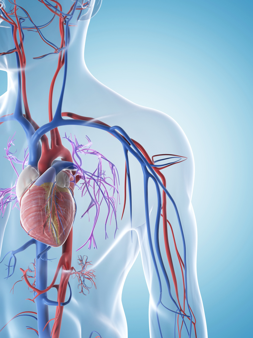
Image 15: Vascular System
Open Image 15 in new tab
Watch Video: Heart and Circulatory System (3:04) to view how blood flows through the heart's chambers and valves at a resting adult heart rate (Beats per minute, BPM = 72).
Go to the Anatomy of the Heart from Wisc-Online and click through the animation to view structures and blood flow.
Arteries of the Heart
Coronary arteries supply blood to the heart muscle. Like all other tissues in the body, the heart muscle needs oxygen-rich blood to function. The image below shows the arteries of the heart (coronary arteries).
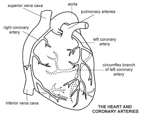
Image 16: Heart and Coronary Arteries
Open Image 16 in new tab
There are 2 main arteries supplying blood to the heart muscle itself:
- Right Coronary Artery (RCA) – supplies blood to right side of the heart
- Left Main Coronary Artery (LMCA) – supplies blood to the left side of the heart
- Left Anterior Descending (LAD)
- Circumflex
Coronary Arteries
Since coronary arteries deliver blood to the heart muscle, any coronary artery disorder or disease can have serious implications by reducing the flow of oxygen and nutrients to the heart muscle.
This reduction in oxygen can lead to a heart attack (also known as myocardial infarction) and possibly death.
Atherosclerosis, or a buildup of plaque in the inner lining of an artery causing it to narrow or become blocked, is the most common cause of heart disease.
This image shows the coronary arteries injected with a dye for visualization using fluoroscopy (a special type of X-ray). Any blockages would be visible using this test.
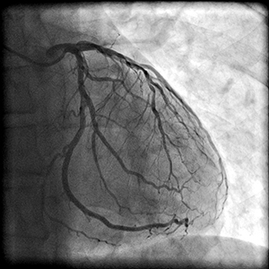
Image 17: Coronary Artery Test
Open Image 17 in new tab
Plaque Build Up
This image shows a comparison of coronary artery lumens.
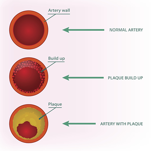
Image 18: Plaque Build Up
Open Image 18 in new tab
Limiting Blood Flow
This image illustrates the narrowing of the artery lumen due to plaque and limiting blood flow. If complete blockage occurs, a heart attack will be the result.
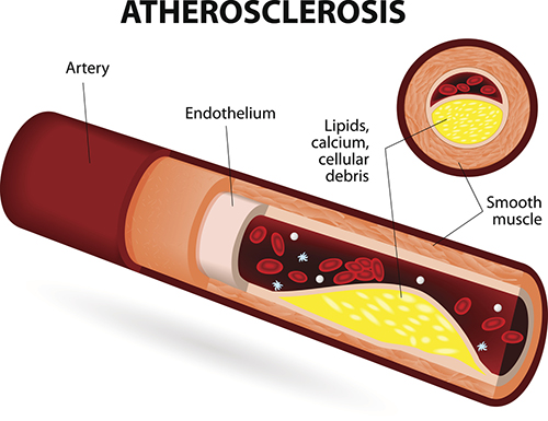
Image 19: Atherosclerosis
Open Image 19 in new tab
![]() Check your understanding...
Check your understanding...
What is your cardiac output if your heart rate is 80 bpm and your stroke volume is 70 ml?
If your heart rate increases, what will happen to your cardiac output?
The main arteries supplying blood to the heart muscle are the ___________ and the __________.