Introduction
- Learn
- Try It
- Task
- Unit Vocab
- Course Vocab
Introduction
What do these images have in common?
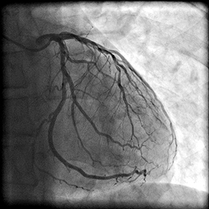
Image 1
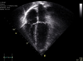
Image 2
These are both images of the heart. Each image is a represenation of the "plumbing" and "mechanical" systems of the heart.
- The first image (Image 1) shows the coronary arteries of the heart ("plumbing system"). These are the vessels supplying blood to the heart muscle. If one gets blocked, a heart attack occurs! Heart attacks are the #1 cause of death in men and women worldwide.
- The second image (Image 2) is an echocardiogram of a beating/moving heart ("mechanical system"). This image is produced with sound waves and shows the chambers and structures of the heart.
Images are important because they can tell you if someone is having a heart attack or having problems with heart valves.
Now, let's learn more about the heart and its plumbing and mechanical systems.
| Lesson Objectives |
|
Following successful completion of this lesson, students will be able to...
Enduring Understandings
The above objectives correspond with the Alabama Course of Study:Anatomy and Physiology standards: 7a and 7b |
![]()