Sarah Smith works as a veterinarian. One evening, she had returned to work for an emergency when she heard a disturbance in the office. She followed the noise to the examination room that contains the medicine cabinet. A woman with dark hair wearing a dark jacket covered in animal fur turned from the cabinet, pushed past Sarah roughly, and exited the front door. She ran off through a neighboring wooded area. Sarah did not pursue her.
Upon inventory of the medication, Sarah found that thousands of dollars of heartworm medication were missing, along with some stronger medication that could be harmful to humans. Sarah immediately called the police.
The Victim
Police reviewed the security camera footage, but could not get a good view of the burglar's face due to her hood and hair. The police also questioned Sarah.
Sarah: She was a little taller than me, maybe 5'6" or 5'7". She was wearing gloves and a neck gaiter, too, so I didn't see much, except her hair and her eyes. Her hair was shoulder-length, maybe longer? It was brown; a lot like mine. Her hoodie was covered with fur. She looked familiar.
The Evidence
Police dusted the medication room for fingerprints, but did not find any. The burglar did not leave any clear footprints. The only clear evidence from the room was hair swept up from the tile floor.
The Suspects – Mary Brown
After speaking to Sarah, police narrowed down the suspects to three people.
Suspect 1 is Mary Brown, former partner at the clinic. Mary was fired the previous year after giving away expensive medication to friends. Mary has pet cats and dogs.
Mary: You want a hair sample? Sure, why not. But I would never set foot back in that place anyhow.
The Suspects – Kate Krebs
Suspect 2 is Kate Krebs. Kate has no known connection to the vet clinic. However, her husband had been caught and jailed for a series of similar burglaries at other local animal hospitals. Kate has pet cats, but is allergic to dogs.
Kate: Burglary? That's not me, that's my husband. I'm not into that kind of stuff. I'll be happy to give you a hair sample if it gets you off my back.
The Suspects – Ashley Hicks
Suspect 3 is Ashley Hicks. Ashley works with a local dog rescue. Last week she came to the clinic asking for donations of heartworm medication and treatment for their rescues. Sarah, concerned about the crowding situation at the rescue, only offered to help if some dogs were boarded at the vet clinic. Ashley was furious and left in a rage.
Ashley: Sarah Smith got robbed? Oh that's rich. Sure, take a hair sample. I doubt you will find anything in that mess of a clinic anyhow. And she was concerned about the condition of MY rescue?
The Forensic Lab
You're a forensic scientist in the crime lab. The detectives on this case have asked you to determine if there is any potential evidence in the hair samples they collected at the crime scene.
They are not very optimistic, since the scale used to weigh animals is also in the medication room. They are concerned that all of the samples are fur, not hair.
Examination Goals
If you do find a human sample, police may be able to perform DNA analysis.
You have two goals in your examination:
Analyze the collected samples and determine if they are human.
Examine any human hair found to determine if it is consistent with the samples from any of the suspects.
Background
Hair Structure
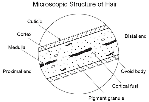
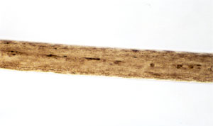
As you know, a hair shaft has three basic regions. Examine the hair shaft below to review.
Cuticle: The cuticle is a protective coating made of overlapping scales. These scales always point toward the tip of the hair. Examination of the cuticle allows investigators to know if the hair is human or not.
Cortex: The cortex gives hair its color and shape. There are three main features found in the cortex—melanin (the pigment that gives hair its color), cortical, and ovoid bodies.
Medulla: The center of the hair, known as the medulla, is surrounded by the cortex. Not all hairs have a medulla. Animal hair can have special types of medulla patterns not found in humans. These distinct medulla patterns help to differentiate between human and animal hairs if there is question about the origin of the hair. It is important to note that medullae are only found in animal hair (including humans) and are never found in plant fibers!
Differentiating Hair
You can differentiate between hair samples in several ways.
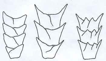
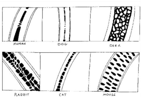
Scale Patterns: You can examine the scale patterns of the cuticle to determine if the hair is human or not. There are three possible scale patterns present on the cuticle:
coronal (mice and rodents)
spinous (cats)
imbricate (humans and dogs)
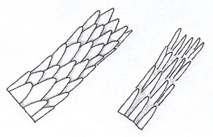
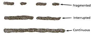
Medulla Patterns: Not all human hair contains a medulla. However, there are three basic medulla types found in human hair:
fragmented
interrupted
continuous
By identifying a medulla type, you may determine if a hair sample is consistent with a person.
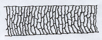
Medulla Structure: The patterns within the medulla can also help distinguish what species hair is from.
Roots: The root of a hair can also be used to determine if a hair is animal or human.
cat
dog
human
Other Features: Scientists can also examine hair for pigment granules, cortical fusi, and ovoid bodies. These patterns may help identify hair as human or animal and match samples.
Examining Hair
Hair can be examined using two different procedures:
Scale Cast: Forensic scientists create an impression (or cast) of the cuticle. They can then study the scale pattern left in the cast to determine the species.
Whole Mount: Scientists can mount the hair samples on a slide. This allows the scientists to examine many features of the hair, including medulla patterns and structures, roots, pigment granules, and ovoid bodies. Examination of the whole mount can help determine the species and identify similar characteristics between hair samples.
Knowledge Check #1
Which of the following characteristics can help you determine the species of a hair sample?
scale pattern
medulla structure
root
all of these
Answer: d. all of these. All of these traits can help determine the species of a hair sample.
Knowledge Check #2
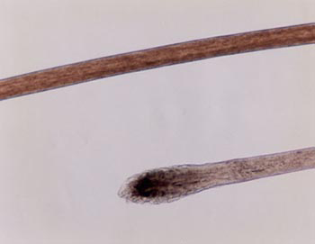
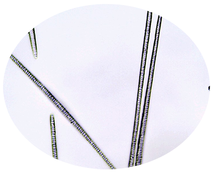
Which species is this hair from? (Hint: Look at the medulla.)
dog
human
cat
Answer: b. human. You can tell based on the fragmented medulla. The cortex is also large relative to the medulla. Finally, the pigment is distributed towards the cuticle.
Knowledge Check #3
Why do you need to collect a sample of hair from Sarah?
Answer: You need to have her hair for comparison since she was also in the examination room with the medication cabinet.
Your Job
You will create a scale cast to determine the species of the hair samples. You will then compare the human evidence samples with samples from the suspect and victim to determine if DNA analysis is needed.
Reflection
What is the purpose of completing a scale cast analysis before examining the hair using a microscope?
Lab Procedure - Species ID Scale Castings
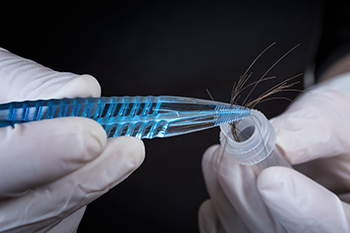
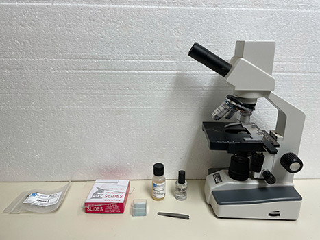
The Equipment
You will use the following equipment to examine the hair evidence.
From left to right: hair samples, microscope slides, cover slips, mounting medium, clear nail polish, forceps, and microscope.
Hair Samples: The hair samples are kept in separate evidence bags to avoid contamination. You have hair from the crime scene plus hair samples from the suspects and Sarah Smith, the veterinarian.
Microscope Slides: You will use the slides to create scale casts and for the whole mounts of the hair.
Cover Slips: The cover slip protects the microscope and holds the sample in place.
Mounting Medium: The mounting medium bonds the slide, sample, and coverslip together. It helps hold the sample in place.
Clear Nail Polish: Clear nail polish is the medium we will use to create our scale castings.
Forceps: You can use forceps (or tweezers) to handle and place the evidence.
Microscope: The microscope will magnify the objects up to 40 times, allowing you to observe tiny differences in the evidence.
Procedure - Scale Castings
Label the slides A, B, and C.
Paint a stripe of clear nail polish onto a microscope slide.
Place the appropriate hair sample on each slide with a short end of the hair extending beyond the edge of the slide. Allow the polish to dry.
Remove the hair samples from the slides by pulling upward on the extended ends of the hairs.
Place each slide under a microscope. Scan the scale patterns using the 4x objective (40x combined magnification), then view the samples under the 10x objective (100x combined magnification), and 40x objective (400x combined magnification).
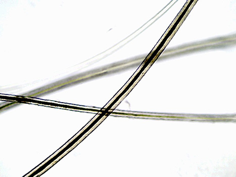
Evidence - Scale Castings
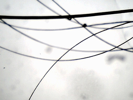
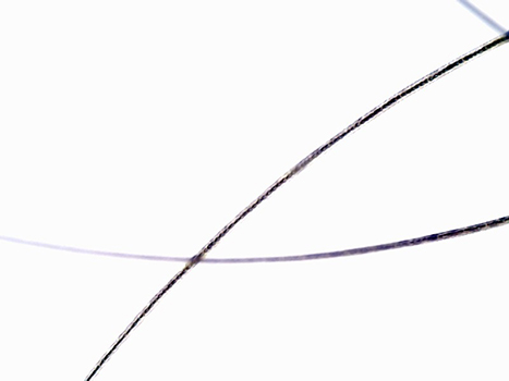
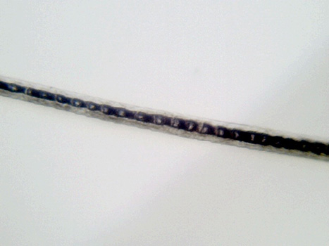
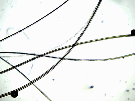
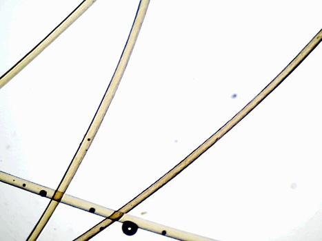
Now that you have made your scale castings, let's see what clue they give to the species present. We know we could have cat, dog, or human hair.
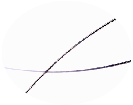
Scale Casting AScale Casting BScale Casting C
Evidence - Species ID Scale Castings
Viewing the Evidence
View all three scale castings below. Sketch what you see in your lab report and make notes on the scale patterns.
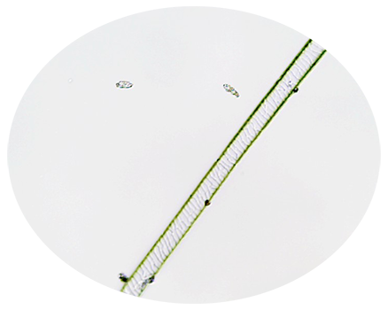
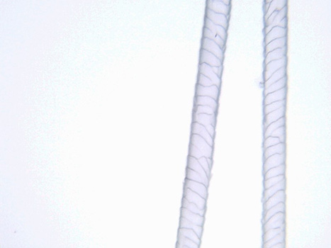
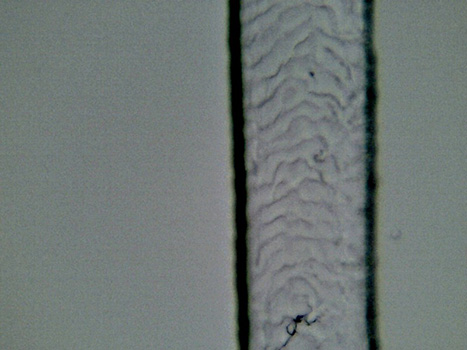
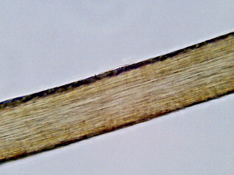
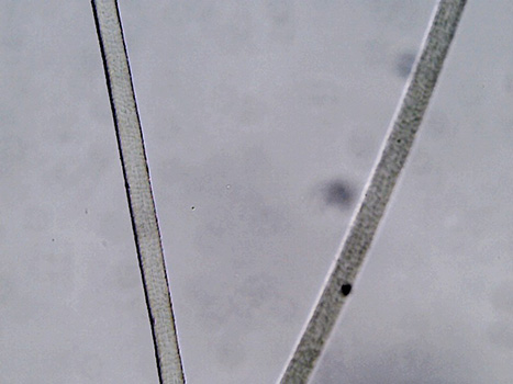
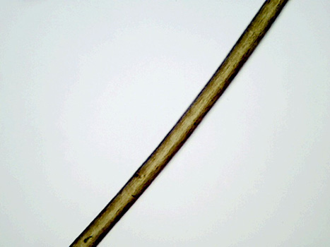
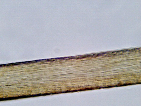
Sample A:
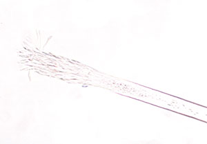
Scale Casting A at 4x magnification.Scale Casting A at 10x magnification.Scale Casting A at 40x magnification. Notice the regular pattern and shape of the scales.
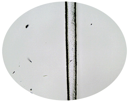
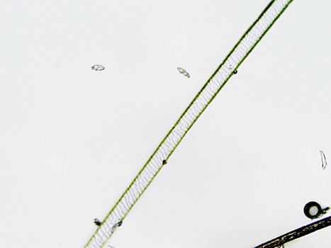
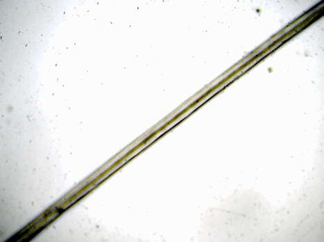
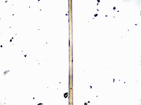
Sample B:
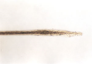
Scale Casting B at 4x magnification.Scale Casting B at 10x magnification.Scale Casting B at 40x magnification.
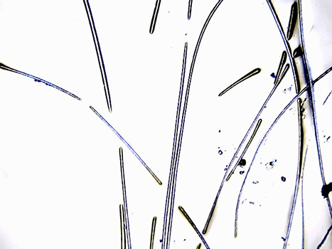
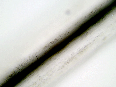
Sample C:
Scale Casting C at 4x magnification.Scale Casting C at 10x magnification.Scale Casting C at 40x magnification.
Lab Procedure - Species ID Whole Mount
Species ID Whole Mounts
You probably were able to tell some differences between the hair samples by studying the scale castings. However, to make definitive species identifications, you will study whole mounts of the hair samples.
You will use the same equipment listed previously in the lab.
Procedure - Species ID Whole Mounts
Place a clean microscope slide on a flat surface. Label the slide.
Select about 5 hairs. Wet a small surface of the slide with the mounting medium. Place the hair sample in the mounting medium to secure the hair in place.
Holding a cover slip horizontally in one hand, add 2 drops of mounting medium to the cover slip.
Quickly invert the cover slip onto the slide starting at one edge and pivoting the other edge down. This helps eliminate trapped air.
View the slide at the lowest magnification (4x objective) on the microscope, then progress down to a higher objective lens magnification (10x magnification). Finally, observe the slides at 40x total magnification.
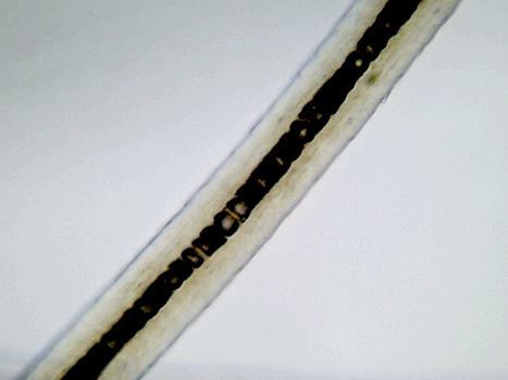
Evidence - Species ID Whole Mount
You now have whole mounts of three different species of hair. You can compare this evidence to the scale castings to make final determinations of species for each collected sample.
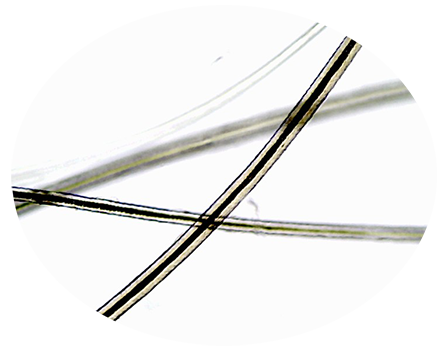
Whole Mount AWhole Mount BWhole Mount C
Evidence - Species ID Whole Mount
Viewing the Evidence
View all three whole mounts below. Sketch what you see in your lab report and make notes of your observations.
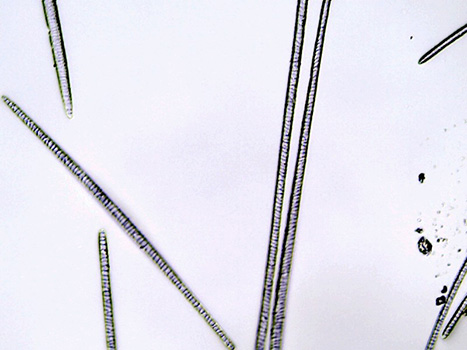
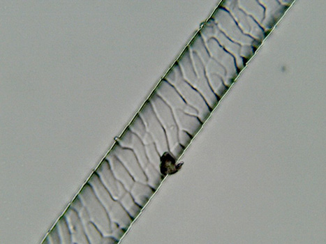
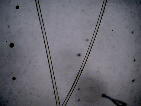
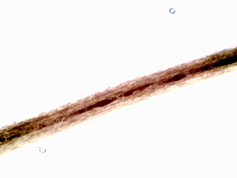
Sample A:
Whole Mount A at 4x magnification.Whole Mount A at 10x magnification.Whole Mount A at 40x magnification.
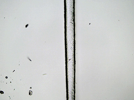
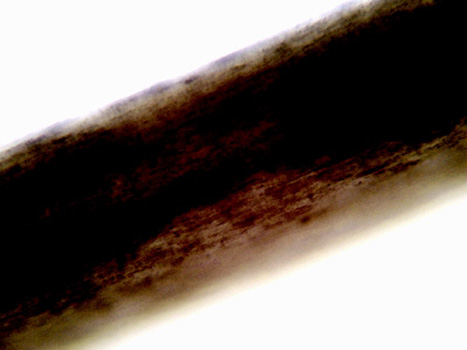
Sample B:
Whole Mount B at 4x magnification.Whole Mount B at 10x magnification.Whole Mount B at 40x magnification.
Sample C:
Whole Mount C at 4x magnification.Whole Mount C at 10x magnification.Whole Mount C at 40x magnification.
Lab Procedure and Evidence - Suspect ID
Suspect Identification
Now that we have identified which one of our hair samples is human, we can finally compare it to the hair samples from Sarah and the suspects.
The mounting process is the same as for the species ID whole mounts, so we will skip directly to the evidence.
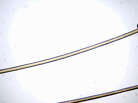
Viewing the Evidence
Observe the evidence from the crime scene. Then, compare it with the samples from the suspects. Record your observations on your lab report.
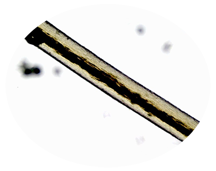
Evidence Sample:
Evidence Sample at 4x magnification.Evidence Sample at 10x magnification.Evidence Sample at 40x magnification.
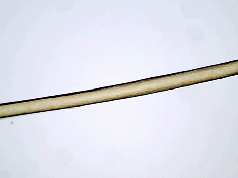
Sarah Smith:
Sarah Smith's hair sample at 4x magnification.Sarah Smith's hair sample at 10x magnification.Sarah Smith's hair sample at 40x magnification.
Mary Brown:
Mary Brown's hair sample at 4x magnification.Mary Brown's hair sample at 10x magnification.Mary Brown's hair sample at 40x magnification.
Kate Krebs:
Kate Krebs's hair sample at 4x magnification.Kate Krebs's hair sample at 10x magnification.Kate Krebs's hair sample at 40x magnification.
Ashley Hicks:
Ashley Hicks's hair sample at 4x magnification.Ashley Hicks's hair sample at 10x magnification.Ashley Hicks's hair sample at 40x magnification.
Analysis
At this point, you should have enough information to identify a suspect. Complete your lab report, being as specific as possible with your observations. Don't forget to include your suspect, if you were able to identify one!
Remember to complete your lab report! Your findings will be shared with law enforcement and used, in part, to resolve or move this case forward.