Learn
Body Cavities
The human body is divided into two portions: the axial and the appendicular. The axial portion includes the head, neck, and trunk while the appendicular portion includes the upper and lower limbs. Body cavities compartmentalize the internal body environment and are lined by membranes. They contain multiple organs within each cavity.
- Below is a list and description of the body cavities along with an illustration. Take time to learn these as you will see them again. You can open a larger version of Image 1 here.
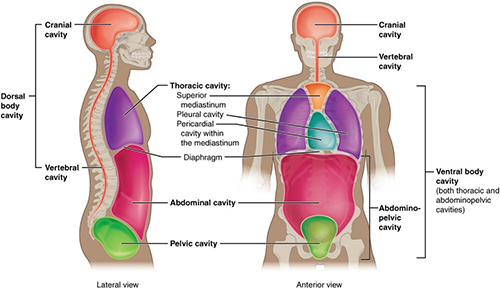
Image labeling the different cavities in a human body. Two views are shown. Image on the left is side facing, or a lateral view. Image on the right is front facing, or an anterior view.
Image 1: Body Cavities
- Dorsal Cavity – protects organs of the nervous system and has two subdivisions:
- Cranial cavity – encloses the brain
- Spinal cavity (also termed vertebral cavity) - encloses the vertebral column and the spinal cord.
- Ventral Cavity – contains subdivisions of the upper and lower trunk:
- Thoracic cavity – surrounded by the ribs and chest muscles. Subdivided into:
- Mediastinum – contains the pericardial cavity
- Pericardial cavity – contains the heart, trachea, and esophagus.
- Pleural cavities – contains the lungs.
- Abdominopelvic cavity – from the diaphragm to the lower floor of the pelvis. Contains two subdivisions:
- Abdominal cavity – includes the liver, stomach, spleen, kidneys, and intestines.
- Pelvic cavity – includes the bladder and reproductive organs.
- Thoracic cavity – surrounded by the ribs and chest muscles. Subdivided into:
Membranes of Body Cavities
The walls of the ventral body cavity and outer covering of its organs contain a thin covering called the serosa (also termed serous membrane).
Serous Fluid: Between the serous membranes is a thin layer of fluid called serous fluid that acts as a lubricant, allowing organs to move within the cavity without causing friction. Serous fluid is secreted by both membranes. In Image 2 you can see where the serous fluid would be by looking at the empty space in the balloon.
Naming of Serous Membranes: The naming of these membranes is done according to the cavity and organ they associate with. An example is the parietal pericardium which lines the pericardial cavity.
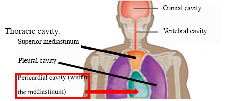
Image highlighting the pericardial cavity. The labels from top to bottom are: cranial cavity, vertebral cavity, and Thoracic cavity. The thoracic cavity includes the superior mediastinum, the pleural cavity, and the pericardial cavity.
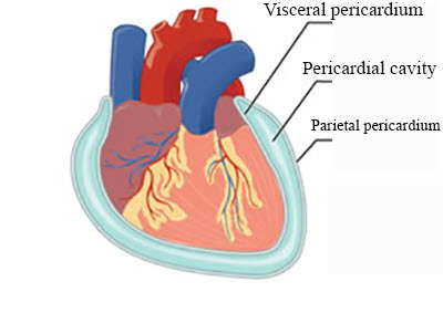
Detail view of the pericardial cavity. Inside of the cavity is a human heart. It is surrounded by the parietal pericardium, the pericardial cavity, and the visceral pericardium.
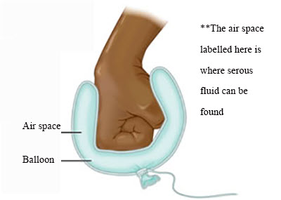
Example of the pericardial cavity. A person's fist represents the human heart. The ballon represents the visceral pericardium and the parietal pericardium. The air in the ballon is where serous fluid can be found and represents the pericardial cavity.
Image 2: Example of Pericardial Body Cavity Membrane
![]() Check for understanding:
Check for understanding:
-What is the purpose of serous fluid and where is it found?
-The serosa that lines the body cavity wall is called the _______ serosa while the membrane lining the organs is the ________ serosa.